Performing Bio-AFM on live cells
This application note discusses the use of atomic force microscopy in biological research, particularly for studying live cells and bacteria, single-cell manipulation, and force spectroscopy.
We highlight the integration of Bio-AFM with optical microscopy and the importance of maintaining physiological conditions during experiments. Learn about the use of various AFM accessories to create a near-physiological environment and manipulate cells. These tools enable precise control over temperature, humidity, pH, and media perfusion, crucial for accurate and relevant biological experiments.

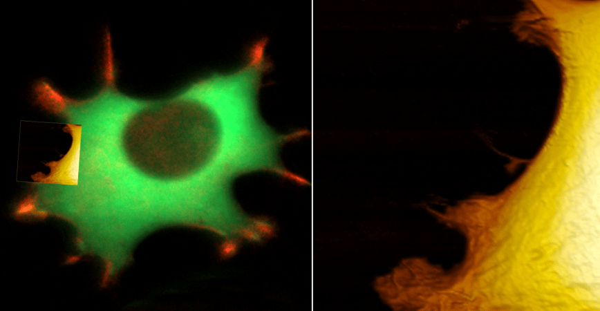
Live fibroblast imaged with inverted light microscope and AFM under cell culture conditions
Left: cell with genetically encoded fluorescent makers (actin-mcherry, gfp-paxilin).
Right, AFM image, 10 μm width.
What you will learn
-
Integrate Bio-AFM with Optical Microscopy: Understand the benefits of combining Bio-AFM with optical microscopy techniques for enhanced biological research.
-
Maintain Physiological Conditions in Experiments: Learn the importance of creating and maintaining physiological conditions, including temperature, humidity, and pH, for live cell experiments.
-
Utilize Accessories for Bio-AFM: Gain knowledge on how to use various accessories like the Live Cell Incubator, Petri dish holder, and perfusion-insert for precise experimental control.
-
Perform Temperature and Humidity Control: Understand the methods to control temperature and humidity in the experimental setup to mimic physiological conditions.
-
Conduct Perfusion of Cell Media: Learn the techniques for controlled disturbance of cells, such as media change or chemical addition, using the perfusion-insert.
-
Explore Single Cell Force Spectroscopy and Cell Manipulation: Discover the applications of single cell force spectroscopy and cell manipulation using FluidFM® in biological research.